Left Hip Muscles Anatomy : Gross anatomic findings after dissection of a left hip in ... : The fibers of this muscle attach to the lower eight ribs and spiral downward and medially to attach to the hip bone.
Left Hip Muscles Anatomy : Gross anatomic findings after dissection of a left hip in ... : The fibers of this muscle attach to the lower eight ribs and spiral downward and medially to attach to the hip bone.. Muscles, connected to bones or internal organs and blood vessels, are in charge for movement. Your email address will not be published. Almost all muscles cross at least one joint (moveable connection between two bones) and cause an action across that joint. Several muscles cross the front of the hip and create hip flexion, pulling the thigh and trunk toward each other, but probably the most important is the iliopsoas. Anatomy of the muscular system.
Microscopic anatomy of skeletal muscle. In human anatomy, the muscles of the hip joint are those muscles that cause movement in the hip. Through a simple and intuitive interface it is possible to observe every anatomical structure from any angle. Rectus femoris muscle, one of the quadriceps muscles on the front of your thigh. In conclusion, a thorough understanding of pelvic and hip anatomy is important for.

If left unstretched, shortened hip flexors affect the position of the pelvis, which in turn affects the position and movement of the lower back.
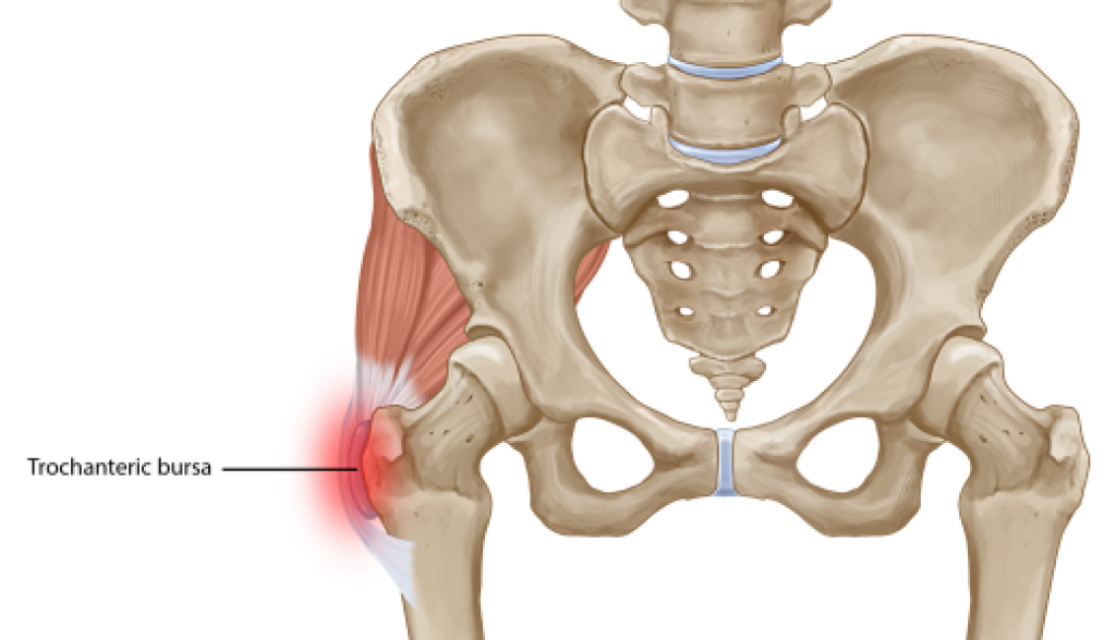
The hip joint is the articulation of the pelvis with the femur, which connects the axial skeleton with the lower extremity. for detailed anatomy of pelvic bones, read anatomy of hip bone. Movement of the femur on the hip in a direction away from the midline of the body in the frontal plane. A bursa that sometimes causes problems in the hip is sandwiched between the bump on the outer hip (the greater trochanter) and the muscles and tendons that cross over the bump. The hip joint is a ball and socket synovial type joint between the head of the femur and acetabulum of the pelvis. Anatomy, bony pelvis and lower limb, psoas major. Almost all muscles cross at least one joint (moveable connection between two bones) and cause an action across that joint. Many doctors, no one believed there was anything wrong. In human anatomy, the muscles of the hip joint are those muscles that cause movement in the hip. Flexion of leg at knee. Several muscles cross the front of the hip and create hip flexion, pulling the thigh and trunk toward each other, but probably the most important is the iliopsoas. Microscopic anatomy of skeletal muscle. The psoas major muscle (usually shortened to just the psoas muscle) is one of the muscles of the posterior abdominal wall and lies not in the retroperitoneum but posterior to it, in the iliopsoas compartment.
It's hard to remember them all! Microscopic anatomy of skeletal muscle. If left unstretched, shortened hip flexors affect the position of the pelvis, which in turn affects the position and movement of the lower back. There are a lot of muscles of the hip and thigh. The hip muscles encompass many muscles of the hip and thigh whose main function is to act on the thigh at the hip joint and stabilize the pelvis.
This anatomical atlas was especially designed for a specific public (radiologists, surgeons, rheumatologists and physicians specializing in musculoskeletal imaging).
Highly detailed 3d models, with textures up to 4k resolution, enable to examine the shape of each. The neck muscles, including the sternocleidomastoid and the trapezius, are responsible for the gross motor movement in the muscular system of the head and neck. Movement of the femur on the hip in a direction away from the midline of the body in the frontal plane. A bursa that sometimes causes problems in the hip is sandwiched between the bump on the outer hip (the greater trochanter) and the muscles and tendons that cross over the bump. This muscle assists with the external rotation of the hip. The function of the quadriceps muscles are: Many doctors, no one believed there was anything wrong. The psoas major muscle (usually shortened to just the psoas muscle) is one of the muscles of the posterior abdominal wall and lies not in the retroperitoneum but posterior to it, in the iliopsoas compartment. Knee assessment and hip mechanics online course: The hip flexors are strong, powerful muscles that can overtake the. These muscles are responsible for hip joint extension (backward movement). Learn their anatomy efficiently and easily using kenhub's muscle anatomy and reference charts! Most modern anatomists define 17 of these muscles, although some additional muscles may sometimes be considered.
A bursa that sometimes causes problems in the hip is sandwiched between the bump on the outer hip (the greater trochanter) and the muscles and tendons that cross over the bump. The function of the quadriceps muscles are: Semimembranosus, semitendinosus and biceps femoris (the hamstrings). Learn about hip muscles human anatomy with free interactive flashcards. In human anatomy, the muscles of the hip joint are those muscles that cause movement in the hip.

3 months later i got acute excrutiating pain in inguinal area.
Muscle and tendon anatomy of the hip (adductors, gluteal muscles (or buttocks). for detailed anatomy of pelvic bones, read anatomy of hip bone. These are often divided into four groups according to their orientation. Anatomy 3d atlas allows you to study human anatomy in an easy and interactive way. Their main function is contractibility. Most modern anatomists define 17 of these muscles, although some additional muscles may sometimes be considered. The hip joint is an intricate structure including hip bones, hip articular cartilage, muscles, ligaments and tendons, and synovial fluid. 936 x 504 png 317 кб. Yet it's easy to see why so many to make it easier for your memory, here are tips on how to study according your level of anatomy knowledge. Knee assessment and hip mechanics learn how hip. Movement of the femur on the hip in a direction away from the midline of the body in the frontal plane. There exist different muscles, which we have covered in class over the past few wee. Through a simple and intuitive interface it is possible to observe every anatomical structure from any angle.


Comments
Post a Comment